Pavilion Publishing and Media Ltd
Blue Sky Offices Shoreham, 25 Cecil Pashley Way, Shoreham-by-Sea, West Sussex, BN43 5FF, UNITED KINGDOM
Tel: +44 (0)1273 434 943
Email: [email protected]


This case study reviews a 69-year-old man who was admitted to a medical ward with the diagnosis of left leg cellulitis and was later diagnosed with leukocytoclasis vasculitis.
A 69-year-old man was admitted to a medical ward with the diagnosis of left leg cellulitis on flucloxacillin. On further questioning, he noticed a rash with blisters on his lower legs two weeks prior to the admission.
Systemic review was unremarkable. Blood results showed:
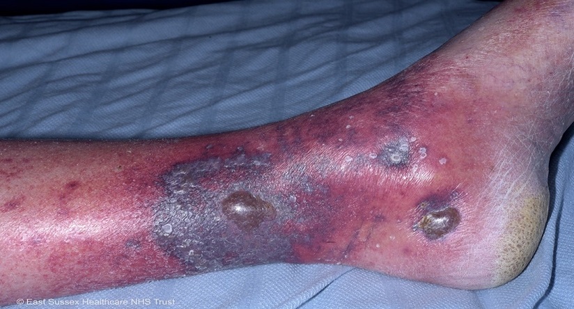
Skin punch biopsy findings were consistent with leukocytoclasis vasculitis. The rash was treated with clobetasol propionate ointment twice daily.
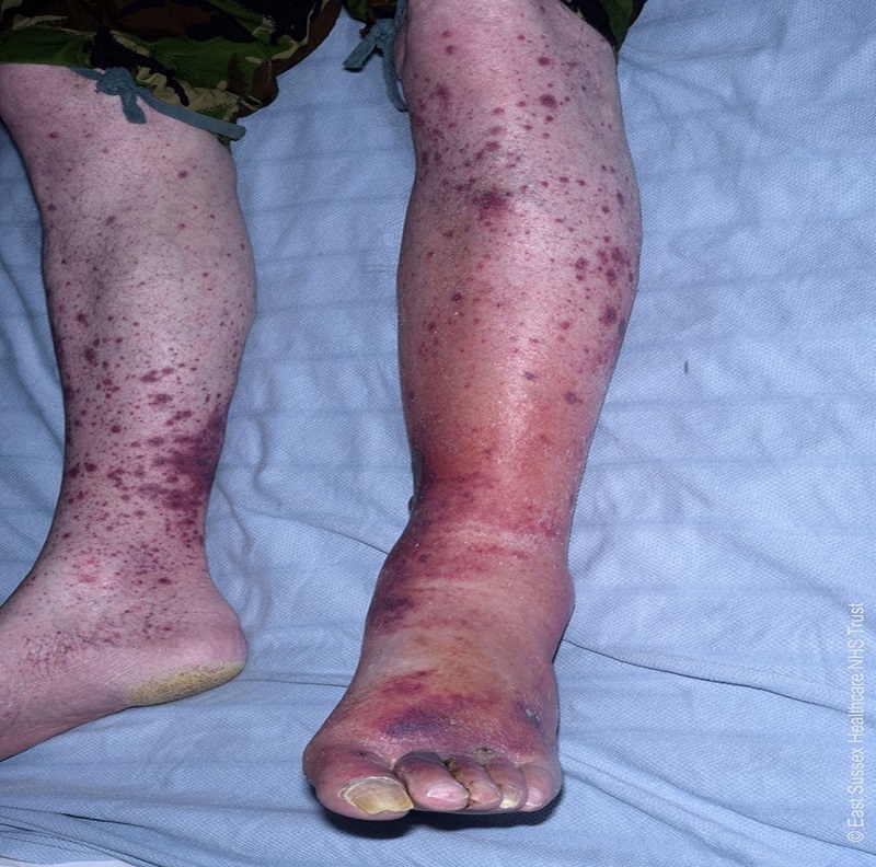
Leukocytoclasis vasculitis is a small vessel vasculitis. Cutaneous leukocytoclasis vasculitis (CLCV) is usually confined to skin with rare extracutaneous manifestations.
The presenting features of CLCV include purpuric rash affecting lower extremities and small vessel involvement.1,2,3
The incidence of biopsy proven CLCV is approximately 45/million individuals/year. If CLCV is suspected, a skin punch biopsy should be performed. Most cases are self-limited with 90% resolving in weeks to months of onset. In persistent vasculitis, treatment depends on the severity of disease and can range from oral corticosteroids to steroid-sparing agents.
Khalesi D Sebastian FY2, Department of Health and Aging, East Sussex Healthcare NHS Trust, St Leonards-on-Sea, East Sussex
Kelum V Ruwanpura, Speciality Registrar, Department of Health and Aging, East Sussex Healthcare NHS Trust, St Leonards-on-Sea, East Sussex
Muhammad JH Rahmani, Consultant Physician, Department of Health and Aging, East Sussex Healthcare NHS Trust, St Leonards-on-Sea, East Sussex

This website uses cookies to improve your experience. We'll assume you're ok with this, but you can opt-out if you wish. Accept Read more ...

