Pavilion Publishing and Media Ltd
Blue Sky Offices Shoreham, 25 Cecil Pashley Way, Shoreham-by-Sea, West Sussex, BN43 5FF, UNITED KINGDOM
Tel: +44 (0)1273 434 943
Email: [email protected]


Müller–Weiss disease is a rare adult-onset osteonecrosis of the navicular bone, typically affecting older women. An 81-year-old presented with six months of left foot pain and swelling, impairing mobility. Radiographs showed a comma-shaped navicular deformity; MRI confirmed collapse and medial resorption. This case highlights the diagnostic value of the “comma sign” and the importance of early recognition to prevent deformity and preserve function.
Müller–Weiss disease (MWD) is a rare cause of chronic foot pain due to adult-onset navicular osteonecrosis. It affects middle-aged or elderly women and often presents insidiously, delaying diagnosis. Radiographs may show navicular collapse, with the “comma sign” indicating lateral deformity.
MRI better visualises necrosis and fragmentation. Management ranges from conservative care to surgery, depending on severity. Early detection is key to avoiding irreversible deformity and functional loss. This case illustrates the clinical and imaging features of MWD in an elderly patient.
An 81-year-old woman presented with a six-month history of persistent pain and swelling in her left foot, accompanied by significant difficulty standing and walking.
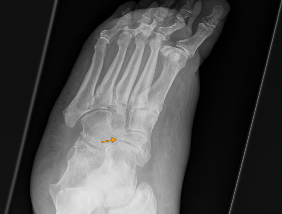
Plain radiography of the left foot revealed a characteristic comma-shaped deformity resulting from collapse of the lateral portion of the navicular bone (Figure 1).
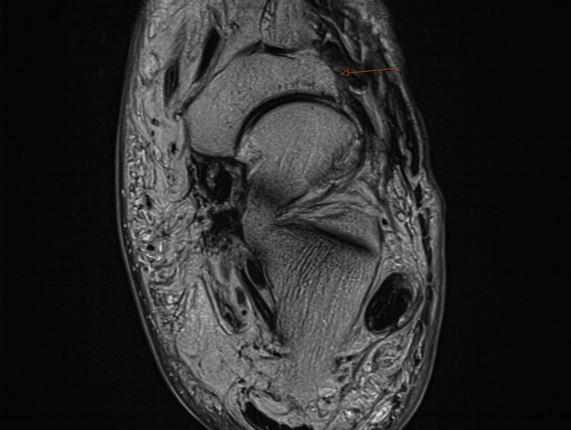
Magnetic resonance imaging (MRI) demonstrated advanced collapse and fragmentation of the navicular, with near-complete medial resorption—findings consistent with Müller–Weiss disease (Figure 2).
Müller–Weiss disease is a rare adult-onset form of avascular necrosis of the navicular bone, most frequently reported in middle-aged or elderly women.1 Clinically, patients present with chronic foot pain, swelling, and progressive deformity. While plain radiographs typically reveal navicular collapse and sclerosis, MRI provides superior visualisation of necrosis and structural compromise.2
Management is guided by disease severity. Conservative approaches include rest, nonsteroidal anti-inflammatory drugs (NSAIDs), and custom orthoses to alleviate symptoms and preserve foot function. In advanced stages, particularly when deformity or pain is pronounced, surgical options such as arthrodesis may be considered.1 Early diagnosis is crucial to prevent further collapse, maintain foot alignment, and preserve mobility. Delayed recognition may result in permanent deformity, impaired ambulation, and reduced quality of life.1,3
Hannah Riches, Care of the Elderly, Conquest hospital, East Sussex Hospital Trust
Gaurav Rajbhandari, Care of the Elderly, Conquest hospital, East Sussex Hospital Trust
Muhammad JH Rahmani, Care of the Elderly, Conquest hospital, East Sussex Hospital Trust

This website uses cookies to improve your experience. We'll assume you're ok with this, but you can opt-out if you wish. Accept Read more ...

