Pavilion Publishing and Media Ltd
Blue Sky Offices Shoreham, 25 Cecil Pashley Way, Shoreham-by-Sea, West Sussex, BN43 5FF, UNITED KINGDOM
Tel: +44 (0)1273 434 943
Email: [email protected]

A case study of a 79-year-old woman who was diagnosed with small cell anaplastic carcinoma after chest x-rays showed a veil sign.
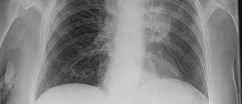
A 79-year-old woman with a history of smoking, recent shortness of breath and weight loss presented to her GP and was started on an empiric course of antibiotic. The anterior posterior (AP) view of the radiograph showed a faint veil like hypolucency covering the left hemithorax with an upward traction of a bulky left hilum and tenting of the left hemi-diaphragm.
The appearance was consistent with left upper lobe collapse presenting as veil sign on the radiograph (Figure 1) Her hospital admission was delayed due to multiple failed attempts at antibiotic courses.
Figure.1 Showing bulky left hilum with tenting of the left hemi-diaphragm and a veil-like opacification covering the left lung due to left upper lobe collapse
Computed Tomography (CT) scan of chest showed a left hilar mass extending in the mediastinum with left upper lobs collapse most likely due to a primary lung malignancy (Figure. 2)
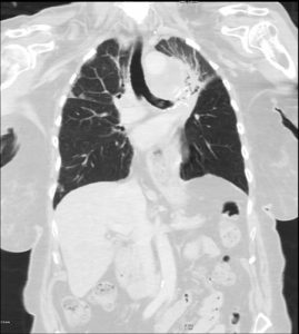
Figure. 2 Left hilar mass (red arrow) extending into the mediastinum and causing left upper lobe collapse (black arrow), most likely due to a primary lung malignancy
Small cell anaplastic carcinoma was diagnosed on bronchoscopic biopsy.
The veil sign develops when the upper lung lobe collapses anteriorly, becoming a thin sheet of tissue opposed to the chest wall and appears as a veiling opacity indicating upper lung lobe collapse.1
The veil sign is not specific for lung cancer – as any cause of left upper lobe collapse may present radiologically in this way but awareness of the importance of veil sign on chest radiograph should lead to prompt referral for investigations to exclude underlying lung cancer in older patients.
Mohammad Farez Sayfoo Foundation Year 1
Amr M. Elyasaky Specialty Registrar
Muhammad JH Rahmani Consultant Physician,
Department of Health and Ageing, Conquest Hospital, East Sussex Healthcare Trust, St Leonards on Sea, East Sussex
Correspondence to Dr MJH Rahmani ([email protected]).

This website uses cookies to improve your experience. We'll assume you're ok with this, but you can opt-out if you wish. Accept Read more ...